How we Take Pictures of the Brain
In the LEAD lab, we use several different methods to understand how your brain learns and grows.
Functional Near-Infrared Spectroscopy (fNIRS)
fNIRS is a brain-imaging technique that lets us measure brain activity. It has several sensors called "optodes" that are arranged inside a stretchy cap, like a swim cap, that we place on your head. Some of the optodes shine small beams of light into your head, and then some of this light reflects back out to the other optodes. How much light is reflected tells us about the blood flow in your brain, which tells us how hard your brain is working! fNIRS is safe, non-invasive, and very kid friendly -- it is small and quiet, and even lets you move around!
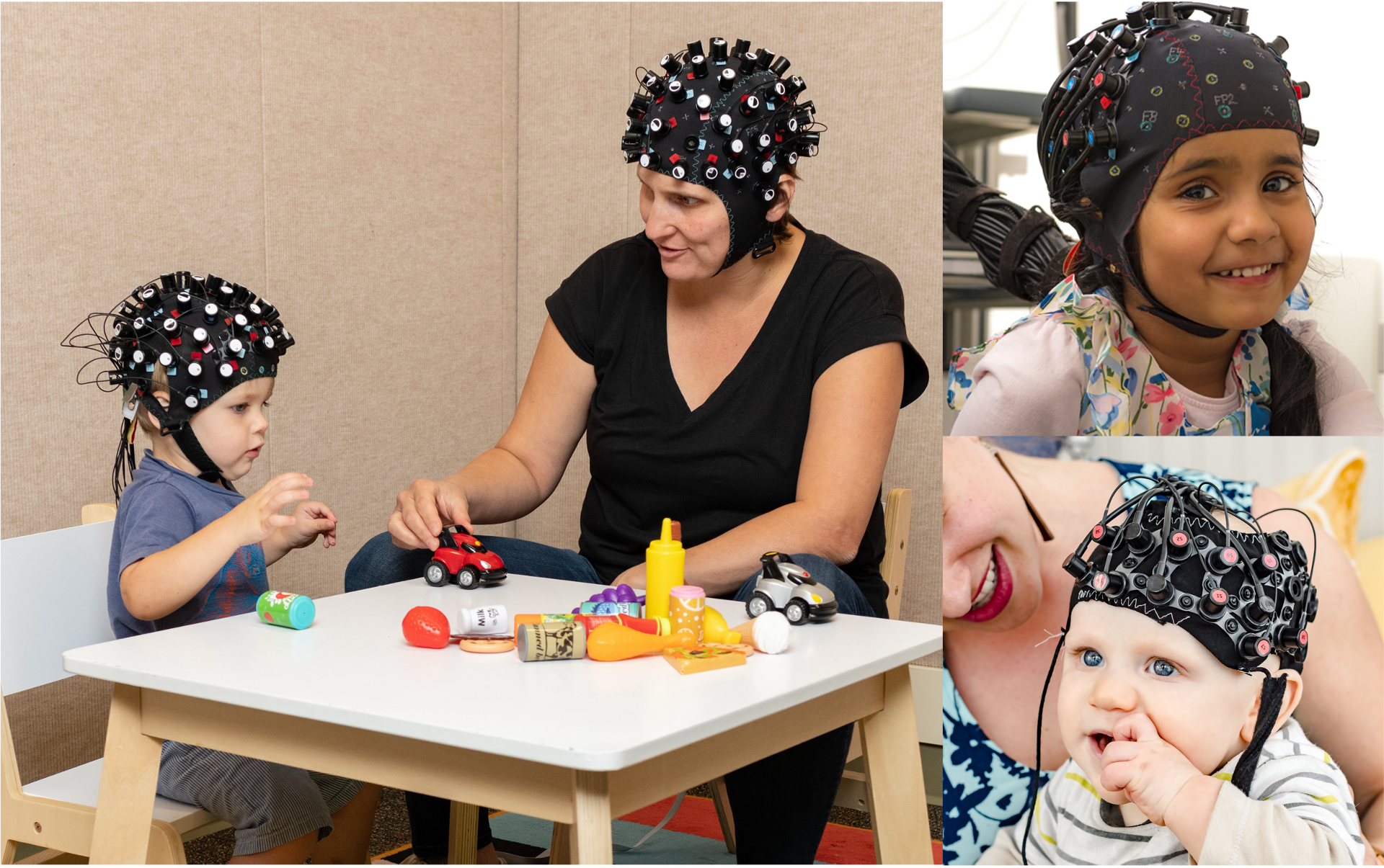
The light emitted from the cap is affected by hair texture, type and color. Specifically, darker and thicker hair can make it harder for the light to reach the brain and accurately measure brain activity. The LEAD Lab has our very own hair equity team who offer to braid participants’ Afro-textured or thick and dark hair to make sure everyone can be included in our research. This allows the fNIRS cap to sit more comfortably on participants’ heads, and enables the light from the cap to reach their brains more easily. Learn more about this process in this video:
Functional Magnetic Resonance Imaging (fMRI)
fMRI is a brain-imaging technique that lets us measure both brain structure and function. MRI uses magnets to track blood flow in your brain, which tells us which parts of your brain you are using while you watch videos, listen to stories, and play games. The magnetic fields can also tell us about other properties of different brain tissues. Because we have to drown out the magnetic field of the earth, we need a REALLY BIG magnet -- this looks like a big tube that you lay down inside. We think it looks like a spaceship! fMRI is also very safe and non-invasive, and we do not use any contrasts or dyes. And when you're done, you will get a picture of your brain to take home!
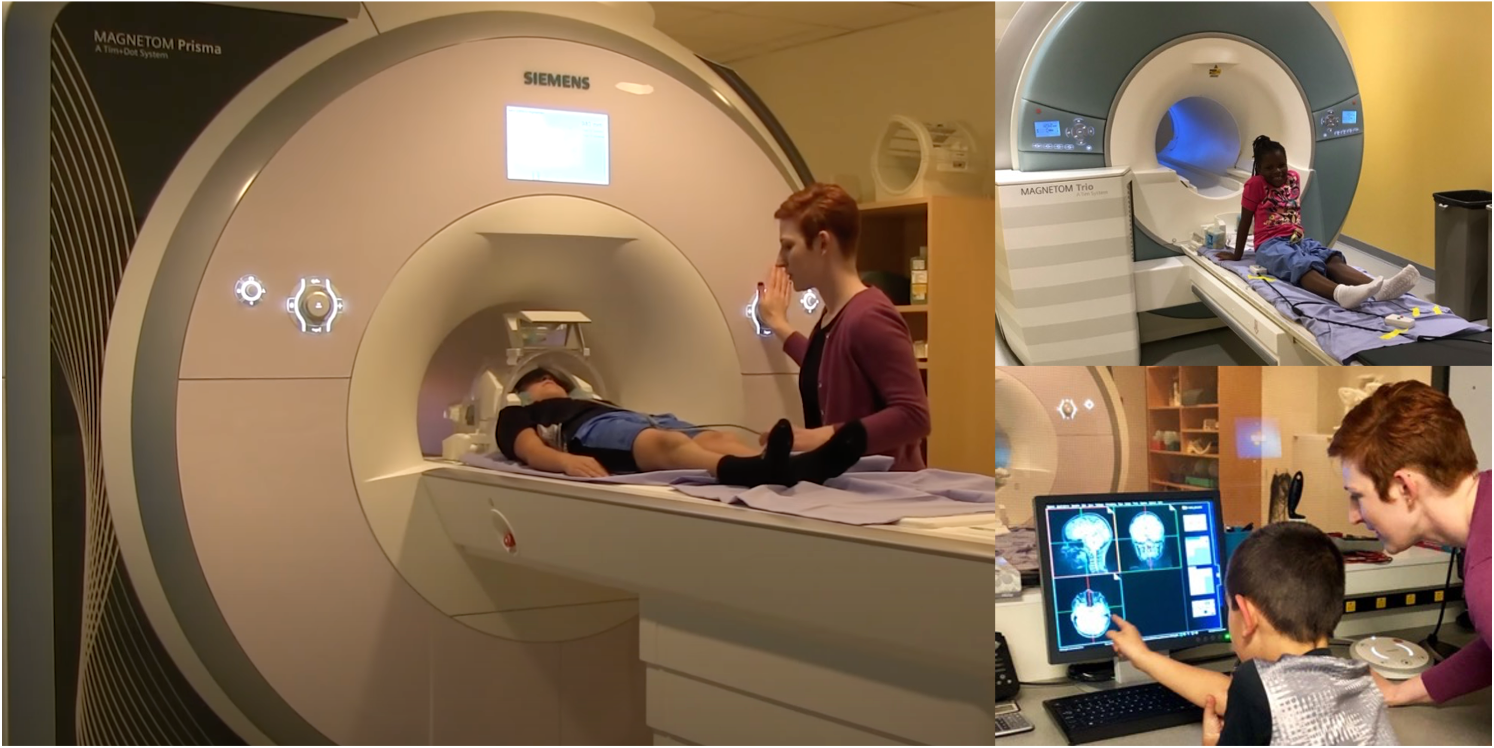
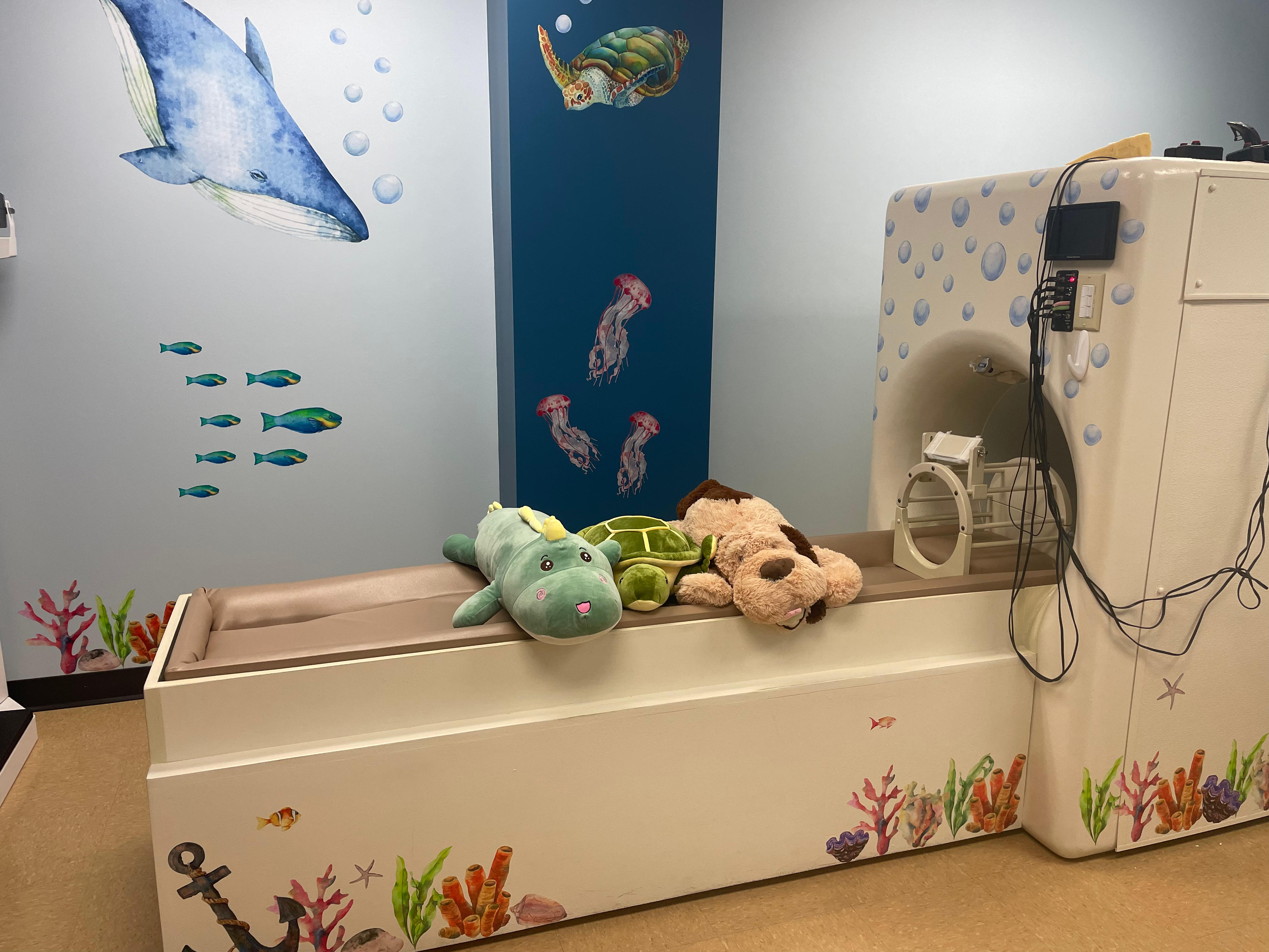
As part of our kid-friendly MRI process, your child will get to snuggle one of our stuffed animal friends during their brain pictures. They're professional brain picture models! Your child can choose to hang out with Churro the dinosaur, Shelley the turtle, or Sir Fredrick the dog during their visit. We conduct MRIs at the Maryland Neuroimaging Center located at 8077 Greenmead Drive, College Park, MD 20740.
Learn more about how MRIs work and how we use them to study the brain by reading the helpful guide below made by the Maryland Neuroimaging Center's DEI Committee.
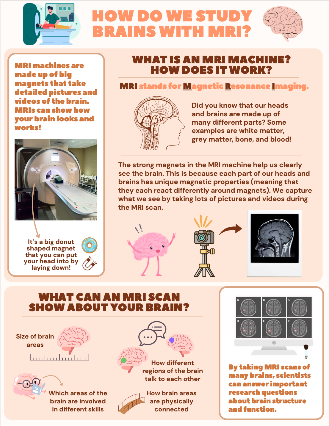
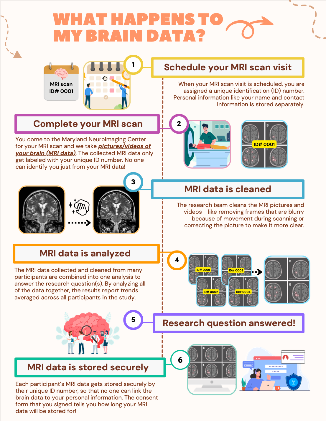
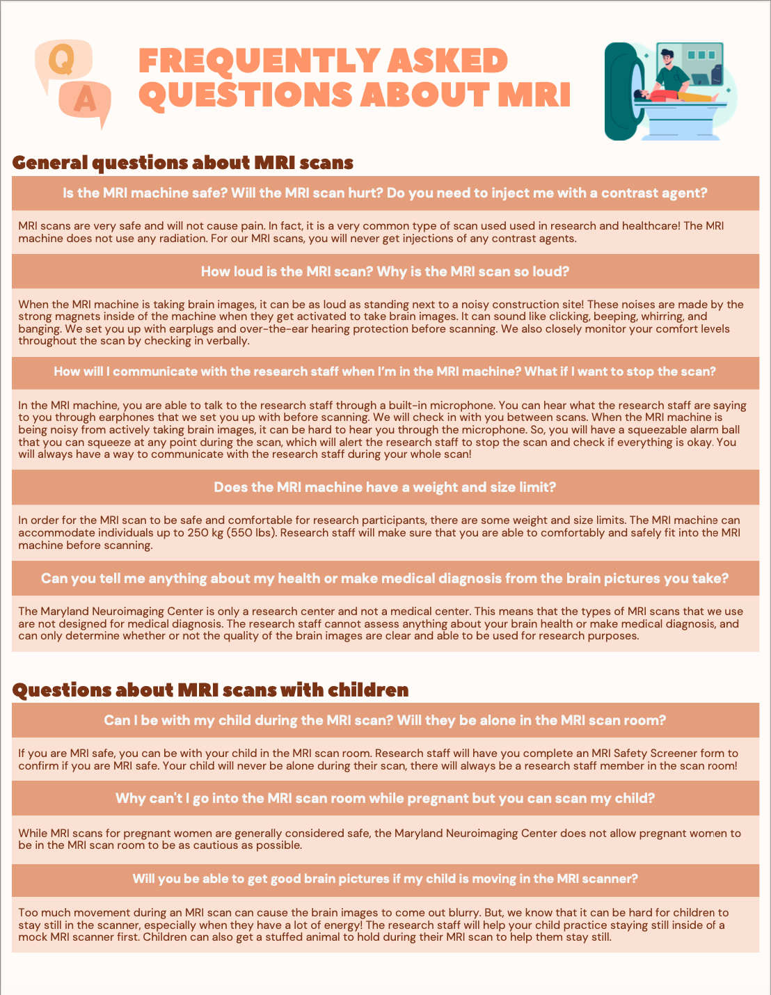
Source: Maryland Neuroimaging Center's Diversity, Equity, & Inclusion Committee